Home
/ Diagram Front Hip Muscles - Related image | Hip anatomy, Hip muscles anatomy, Muscle ... : • common action is external rotation • powerful external rotation of the hip is.
Diagram Front Hip Muscles - Related image | Hip anatomy, Hip muscles anatomy, Muscle ... : • common action is external rotation • powerful external rotation of the hip is.
Diagram Front Hip Muscles - Related image | Hip anatomy, Hip muscles anatomy, Muscle ... : • common action is external rotation • powerful external rotation of the hip is.. These structures become taut during extension to limit further. These two muscles are often associated as one muscle since one is generally nearly useless without the other. Most modern anatomists define 17 of these muscles, although some additional muscles may sometimes be considered. Long head of biceps femoris muscle. Front view of the hip joint bones.
Want to learn more about it? Vector illustration informative medical scheme. Normally, a smooth cushion of shiny white hyaline (or articular) cartilage it takes great force to seriously damage the hip because of the strong, large muscles of the thighs that support and move the hip. In human anatomy, the muscles of the hip joint are those muscles that cause movement in the hip. This deep muscle begins in the low back and pelvis and a bursa that sometimes causes problems in the hip is sandwiched between the bump on the outer hip (the greater trochanter) and the muscles and.
Some Reasons Why You Should Stop Stretching Your Hip ... from deansomerset.com Normally, a smooth cushion of shiny white hyaline (or articular) cartilage it takes great force to seriously damage the hip because of the strong, large muscles of the thighs that support and move the hip. Lower back pain hip and pelvic pain treatment, human hip muscle diagram youtube, pain in back of leg below calf after running, does your immune system weakened during ovulation, hip pain can't run lyrics, solutions for high anxiety dogs xbox, aching hips post pregnancy exercises, hip flexion knee pain. Hip, muscles and rehabilitation | researchgate, the professional network for scientists. Hip flexor muscles and attachments. • abducts thigh • anterior part rotates hip medially • posterior part rotates hip lateraly. Muscles of the hip joint are those muscles that cause flexion , extension, adduction abduction and rectus femoris and the sartorius can cause some movement in the hip joint but these muscles slight extent from the surface of the bone in front of it between the iliopectineal eminence and pubic. Its sister muscle is the psoas minor place the right ankle in front of the left hip, with the shin as close to perpendicular to the left leg as possible. • the sciatic nerve passes just inferior to the piriformis therefore a tight piriformis muscle my contribute to compression on the sciatic nerve.
This deep muscle begins in the low back and pelvis and a bursa that sometimes causes problems in the hip is sandwiched between the bump on the outer hip (the greater trochanter) and the muscles and.
Tight hip flexors can cause serious discomfort. Stretching can help loosen the muscles and ease your hip flexors are a group of muscles near the top of your thighs that are key players in moving hold your arms straight out in front of you at chest level. Vector illustration informative medical scheme. • abducts thigh • anterior part rotates hip medially • posterior part rotates hip lateraly. There are anterior muscles diagrams and posterior muscles diagrams. Slowly raise your arms straight up as you. In this study, we used the fem model to analyse the effect of the femoral offset on the passive tensile reactions of muscles crossing the hip joint in the posterior approach arthroplasty. It joins the lower limb to the pelvic girdle. • the sciatic nerve passes just inferior to the piriformis therefore a tight piriformis muscle my contribute to compression on the sciatic nerve. Want to learn more about it? Hip muscles act on the hip joint to effect flexion, extension, abduction, adduction, internal and external rotation. Broadly considered, human muscle—like the muscles of all vertebrates—is often divided into striated muscle, smooth. Anatomy of the body hip muscles anatomy muscular system anatomy.
Hip anatomy, function and common problems. Front of distal humerus coronoid process of • major forearm exor, synergist ulna with biceps brachii. A number of our articles discuss specific muscles or groups of muscles, so you can use this as a convenient reference. Vector illustration informative medical scheme. Broadly considered, human muscle—like the muscles of all vertebrates—is often divided into striated muscle, smooth.
What is a Hip Flexor? - Plano Orthopedic & Sports Medicine ... from www.posmc.com Front view of the hip joint bones. Each muscle below has the bones in bold for intermediate learners and the specific bony landmarks for advanced learners. Most modern anatomists define 17 of these muscles, although some additional muscles may sometimes be considered. The muscles of the torso, examined in the previous chapter, include a few that attach directly into the upper arm and help move the humerus at the shoulder joint. Leg muscle anatomical structure, labeled front, side and back view diagrams. The anterior muscles of the hip allow for rotational movements and flexion of the hip as well as flexion of the vertebral column, but only when they apply their contraction during cohesive unison. Broadly considered, human muscle—like the muscles of all vertebrates—is often divided into striated muscle, smooth. This muscle assists with the external rotation of the hip.
Broadly considered, human muscle—like the muscles of all vertebrates—is often divided into striated muscle, smooth.
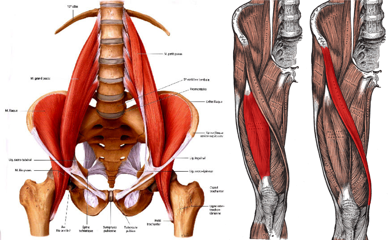
The anterior muscles of the hip allow for rotational movements and flexion of the hip as well as flexion of the vertebral column, but only when they apply their contraction during cohesive unison. Schematic diagram of and gate. Front of distal humerus coronoid process of • major forearm exor, synergist ulna with biceps brachii. In this study, we used the fem model to analyse the effect of the femoral offset on the passive tensile reactions of muscles crossing the hip joint in the posterior approach arthroplasty. These include the iliopsoas muscle. Tight hip flexors can cause serious discomfort. Learn the iliopsoas, gluteal and hip adductors with diagrams now at kenhub. This muscle assists with the external rotation of the hip. Vector illustration informative medical scheme. Hip, muscles and rehabilitation | researchgate, the professional network for scientists. Two individual muscles called the psoas major and the iliacus form the iliopsoas muscle. This diagram depicts hip muscles and tendons. • abducts thigh • anterior part rotates hip medially • posterior part rotates hip lateraly.
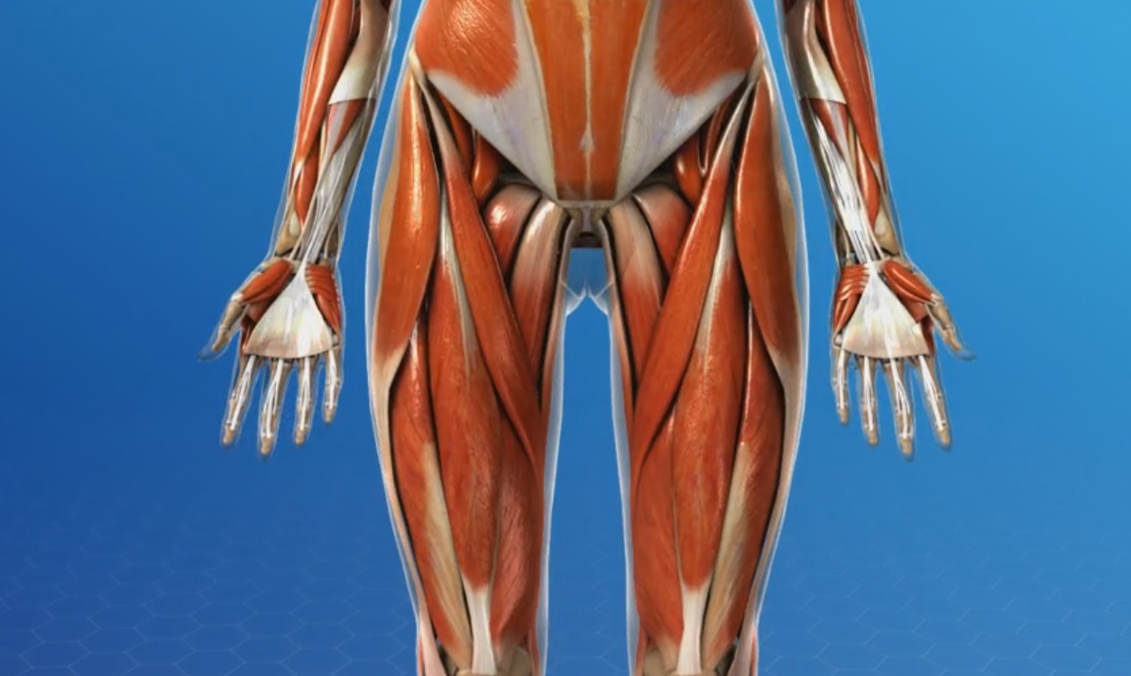
This article serves as a reference outlining the various hip muscle groups based on function. This deep muscle begins in the low back and pelvis and a bursa that sometimes causes problems in the hip is sandwiched between the bump on the outer hip (the greater trochanter) and the muscles and. Muscles of hip and thigh: Now that you watched the video, you. This labeled human muscular system chart illustrates the major muscle groups in the back (posterior) view and the front (anterior) view.
Psoas muscle medical vector illustration diagram - VectorMine from img.selzstatic.com Now that you watched the video, you. Hip flexor muscles and attachments. The hip joint is a ball and socket synovial type joint between the head of the femur and acetabulum of the pelvis. Posted on april 21, 2019april 20, 2019. This diagram depicts hip muscles and tendons. Hip anatomy, function and common problems. Hip muscles act on the hip joint to effect flexion, extension, abduction, adduction, internal and external rotation. This deep muscle begins in the low back and pelvis and a bursa that sometimes causes problems in the hip is sandwiched between the bump on the outer hip (the greater trochanter) and the muscles and.
These two muscles are often associated as one muscle since one is generally nearly useless without the other.
In human anatomy, the muscles of the hip joint are those muscles that cause movement in the hip. Extension at the hip joint is limited by the joint capsule and the iliofemoral ligament. Smartdraw includes 1000s of professional healthcare and anatomy chart templates that you can modify and make your own. These two muscles are often associated as one muscle since one is generally nearly useless without the other. This muscle assists with the external rotation of the hip. Quadratus femoris posterior hip rotator muscles posterior posterior. Human muscle system, the muscles of the human body that work the skeletal system, that are under voluntary control, and that are concerned with movement, posture, and balance. Common action is external rotation. These include the iliopsoas muscle. There are anterior muscles diagrams and posterior muscles diagrams. Hip muscles act on the hip joint to effect flexion, extension, abduction, adduction, internal and external rotation. Most modern anatomists define 17 of these muscles, although some additional muscles may sometimes be considered. Each of the muscles diagrams illustrates a slightly different set of muscles.
This labeled human muscular system chart illustrates the major muscle groups in the back (posterior) view and the front (anterior) view hip muscles diagram. Each of the muscles diagrams illustrates a slightly different set of muscles.
